This poster presents a study on the morphology of the primary cilium in neurons derived from induced pluripotent stem cells (iPSCs) of patients with spinocerebellar ataxia type 17 (SCA17). The researchers aimed to generate brain organoids from patient-derived iPSCs to study primary cilium structures. They used dermal fibroblasts from a healthy donor, a carrier, and an SCA17 patient to induce pluripotent stem cells, which were then differentiated into neuroepithelium, neuronal progenitor cells, and midbrain organoids using specific small molecules and growth factors. The samples were analyzed via 2D culture generation, confocal microscopy, quantitative RT-PCR, and transmission electron microscopy. The results show that the nuclear area in patient cells was 1.5 times smaller than in healthy controls. Additionally, the expression of primary cilia genes DYNC2H1 and TRAF3IP1 in patient cells was significantly decreased by 2.5 and 7.9 times, respectively. Transmission electron microscopy demonstrated that the ultrastructure of the primary cilia in patient organoid cells remained normal. However, immunocytochemical analysis of 2D cultures revealed that the cilia length in patient cells was 1.5 times shorter compared to healthy control cells. In conclusion, brain organoids from both healthy donors and SCA17 patients express predominantly neuronal markers. Although the primary cilia in neurons from SCA17 patients display normal ultrastructure, their length is significantly reduced, and the expression levels of the primary cilia-related genes DYNC2H1 and TRAF3IP1 are substantially downregulated.
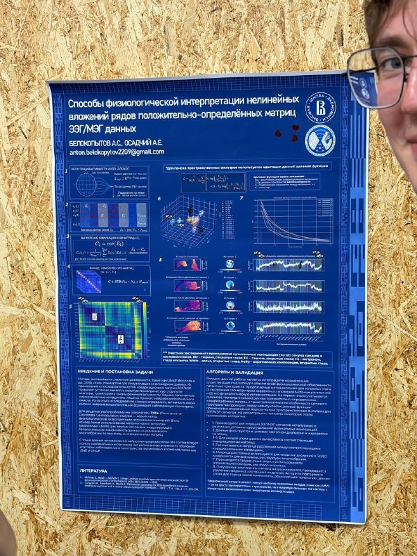
This poster presents research by A. S. Belokopytov and A. E. Osadchiy from the HSE University Center for Bioelectric Interfaces regarding methods for the physiological interpretation of non-linear embeddings in electroencephalography (EEG) and magnetoencephalography (MEG) data. While non-linear dimensionality reduction techniques like UMAP and t-SNE effectively reveal geometric patterns and clusters within high-dimensional covariance matrices, the resulting low-dimensional coordinates generally lack direct physiological meaning, making it difficult to pinpoint which neural populations generate the observed structures. To solve this issue, the authors introduce TriCo (Covariance-to-Coordinate Co-modulation analysis), a method designed to make non-linear latent spaces physiologically explainable. The technique adapts cross-entropy loss optimization on the Riemannian manifold of symmetric positive-definite matrices to find spatial filters and map topological variability directly back to cortical sources. The algorithm was validated using EEG and MEG data recorded while participants listened to 120-second audio segments under various conditions, including resting states with eyes open or closed, metronome beats, waltzes, and non-rhythmic musical compositions. The procedure filters signal data into target frequency bands, epoch-wise covariance matrices are calculated, and pairwise distances are measured to project data into two- or three-dimensional spaces. By iteratively applying gradient descent optimization and extracting specific source components, the algorithm allows researchers to isolate and remove individual source contributions to re-evaluate remaining data topology. Overall, the proposed approach overcomes a major limitation of standard non-linear dimensionality reduction by explicitly connecting abstract clusters in low-dimensional visual space to specific physiological generators of brain activity.
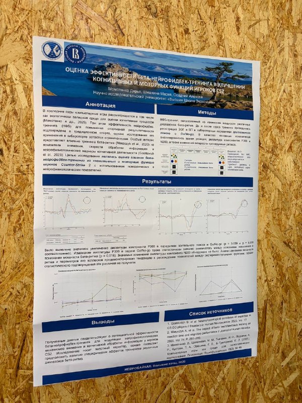
This study evaluates the effectiveness of beta-neurofeedback training in improving the cognitive and motor functions of Counter-Strike 2 players. Conducted at the HSE University, the research examined how altering beta-rhythm power influences behavioral and neurophysiological markers such as reaction times and event-related potential components, specifically P300 and N200, during visual search and Go/No-go tasks. The experiment involved ten right-handed adult participants with active CS2 playing experience, split evenly into beta-upregulation and beta-downregulation training groups. Results showed a statistically significant increase in P300 amplitude following training across both visual search and Go/No-go tasks, as well as a significant difference in P300 amplitude changes between the upregulation and downregulation conditions. No significant changes were observed in the N200 component, and while trends in beta-rhythm power dynamics differed across groups, they did not achieve statistical significance. Ultimately, the pilot findings suggest that beta-neurofeedback training holds potential for modulating the neurophysiological mechanisms underlying attention and cognitive processing in esports players, with different beta-rhythm training protocols yielding distinct effects.
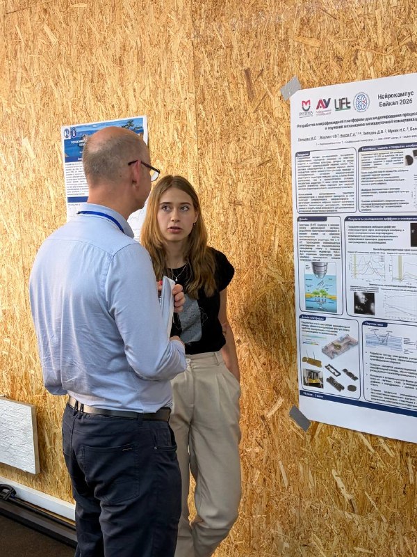
This scientific poster presents the development of a microfluidic platform designed to model synaptic transmission and study intercellular communication mechanisms in vitro. Presented at Neurocampus Baikal 2026 by researchers affiliated with Pirogov Russian National Research Medical University, Alferov University, LIFT Center, and the Federal Center of Brain Research and Neurotechnologies, the project aims to improve preclinical drug testing by creating more accurate cellular microenvironments through the integration of microfluidics and microiontophoresis. The underlying technology relies on a silicon chip equipped with a transparent silicon nitride nanoporous membrane. Coating this membrane with poly-L-lysine maintains electrical conductivity while providing a favorable environment for neural cell adhesion and growth, which was validated using mammalian cells and primary hippocampal cultures. In the test setup, a two-chamber system separates serotonin in the lower chamber from cells expressing serotonin receptors in the upper chamber. Neuromediator transport across the nanopores is actively regulated by applying an external voltage of up to three hundred millivolts, and cellular responses are tracked via fluorescent calcium imaging using Fluo-4. Results confirm both passive diffusion and precise, electrically driven transport, release, and retention of neurotransmitters across the membrane. The authors introduced a user-friendly microfluidic chamber design that simplifies assembly and improves experimental reproducibility. Looking forward, the researchers plan to refine the electrically controlled transport system, implement a closed recirculating perfusion network, validate the platform with additional neurotransmitters such as dopamine, glutamate, and GABA, and build a modular experimental setup offering integrated chemical, optical, and electrical control over cell signaling.
This research poster presented by N. Titova, E. Kuzmina, G. Soghoyan, and M. Lebedev investigates the temporal dynamics of cortical stereotactic EEG activity during the observation and execution of bionic hand gestures. The study re-evaluates previous prominent claims that neural population dynamics are fundamentally rotational, proposing instead that such rotational patterns in multidimensional space are equivalent to neuronal travelling waves or sequences. Experimentally, eight to twelve stereotactic EEG channels and two electromyography channels were recorded at two kilohertz from thirty epilepsy patients as they maintained extended palm positions or flexed fingers matching nine distinct movements of a bionic hand. The results demonstrate that rotational dynamics observed via statistical dimensionality reduction methods correlate directly with the presence of travelling waves across the cortex. Spectral analysis further revealed that slow electroencephalographic rhythms predominate during movement preparation, while gamma and high-gamma rhythms are strongly pronounced during movement execution, mirroring electromyographic activity. Overall, the findings indicate that population-level rotational dynamics can be explained by multichannel travelling waves across specific spectral bands, offering relevant insights for the design and refinement of neuroprosthetic systems.