TGViewer
Channel Public Channel
ECG.CASES

ECG.CASES

@ecgcases

Daily ECG

Admin: @po5eidon

instagram: instagram.com/ecg.cases
Subscribers
12.1K
Photos
424
Videos
0
Links
0

Showing posts older than #925 · Back to latest

Older Posts 20 shown
Post #924 7.55K
  • 👍 4
  • ❤ 2
Post #923 7.54K
  • 👍 6
  • ❤ 4
Post #922 7.08K
In the beginning, there are sinus beats that are conducted with 1st degree AV block. These beats are followed by ectopic atrial contractions which are not conducted through AV node (blocked PACs). However, an ectopic atrial contraction eventually triggers an AVNRT, a regular narrow complex tachycardia with relatively long RP intervals due to slow conduction through retrograde pathway in the AV node.

Special Thanks to my dear friend salvatore salvatore for providing the diagram.
  • 👍 12
  • ❤ 5
Post #921 6.59K
The correct answer is “Atrioventricular nodal reentry tachycardia (AVNRT)”
  • ❤ 1
  • 👍 1
Post #920 8.58K
  • 👍 17
  • 🤯 12
  • ❤ 5
  • 🤔 5
  • 🔥 4
Post #919 8.21K
65 y female with history of heart failure presented with palpitation
  • 👍 11
  • 🤯 8
  • ❤ 5
Post #918 6.7K
#case198
  • 👍 2
  • ❤ 1
Post #917 7.4K
  • 👍 1
Post #916 7.22K
ECG features:
• QRS complexes cluster into groups, separated by short pauses. At the end of each group is a non-conducted P wave (dropped beats)
• The PR interval progressively increases from one complex to the next.
• The P-P interval is relatively constant despite the irregularity of the QRS complexes.
  • 👍 21
  • ❤ 3
Post #915 6.94K
The correct answer is “Second degree AVB: mobitz 1 (Wenckebach)”
  • ❤ 4
  • 👍 1
Post #914 8.08K
  • 👍 13
  • ❤ 10
  • 🤔 3
Post #913 7.57K
55 y man with history of Ischemic heart disease presented with occasional dizziness.
  • 👍 16
  • ❤ 4
Post #912 8.23K
  • 🔥 21
  • 👍 5
  • ❤ 3
Post #911 7.74K
ECG features:
• Inverted flutter waves in Inferior leads at a rate of 350 bpm
• Upright flutter waves in V1 simulating P waves
• 2:1 AV block resulting in a ventricular rate of 175 bpm
  • 👍 14
  • ❤ 1
Post #910 7.49K
The correct answer is “Atrial flutter”
  • 👍 4
  • 🤔 4
  • ❤ 1
Post #909 12.2K
  • 👍 15
  • ❤ 3
Post #908 11.1K
55 y man with history of mitral valve replacement presented with palpitation
  • 👍 16
  • 😁 3
  • ❤ 2
Post #907 10.2K
#case196 ❤️
  • 👍 2
  • ❤ 1
Post #906 10.9K
  • ❤ 7
  • 👍 1
Post #905 10.5K
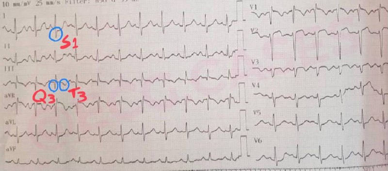
The ECG shows “Sinus tachycardia” and “S1Q3T3 pattern”, two most common ECG findings in PTE. These findings are neither sensitive nor specific for PTE, Thus ECG findings only in combination with physical symptoms suggest PTE.

Here are the most common ECG findings in patients with PTE:

• Sinus tachycardia
• S1Q3T3 pattern
• New-onset atrial fibrillation or flutter
• incomplete right bundle branch block
• right axis deviation
• T-wave inversion in leads V1 to V4
  • ❤ 9
  • 👍 8
Older posts →
Threads Profile ViewerView any public Threads profile without an account.Open ThreadLook →Writing with AI? Make it sound human.Metric37 rewrites AI drafts so they read naturally. Free AI detector, 1,500 words free.Try Metric37 →