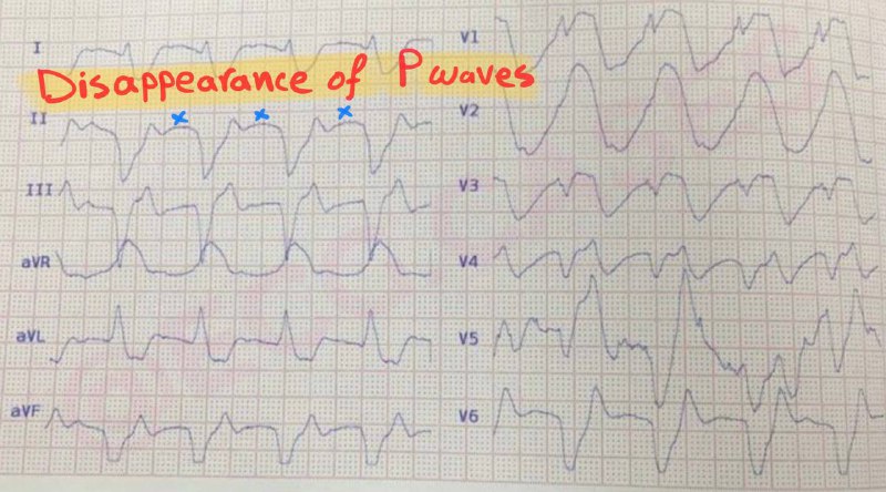
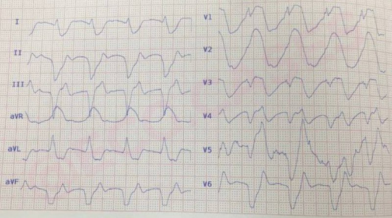
This ECG displays many of the features of hyperkalaemia:
• Disappearance of p waves • ST elevations (best seen in aVR and chest leads) • Broad, bizarre QRS complexes that merge with subsequent ST segment and T wave. • Sine waves (best seen in V3)
There is a regular, broad complex tachycardia at ~187 bpm; All QRS complexes are preceded by P waves with abnormal axis (Negative P in D1). These findings suggest atrial tachycardia. Note that one P wave is dropped in chest leads and made the rhythm irregular.
Other ECG findings: Left anterior hemiblock Ventricular conduction delay
 EC
EC