TGViewer
Channel Public Channel
ECG.CASES

ECG.CASES

@ecgcases

Daily ECG

Admin: @po5eidon

instagram: instagram.com/ecg.cases
Subscribers
12.1K
Photos
424
Videos
0
Links
0

Showing posts older than #818 · Back to latest

Older Posts 20 shown
Post #817 7.38K
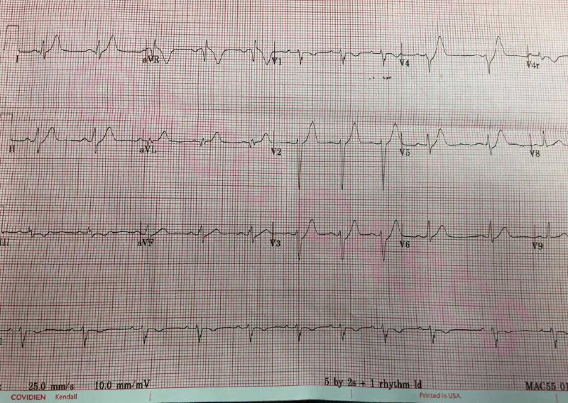
30 y woman presented with chest pain and shortness of breath
  • 👍 3
  • ❤ 1
Post #816 6.58K
#case185 ❤️
  • ❤ 2
  • 👍 1
Post #815 7.14K
  • ❤ 1
  • 👍 1
Post #814 6.6K
  • ❤ 1
  • 👍 1
Post #813 6.1K
#Extra04 ❤️
  • 👍 1
Post #812 7.03K
  • ❤ 1
  • 👍 1
Post #811 6.7K
  • ❤ 1
  • 👍 1
Post #810 6.54K
The ECG shows upslope ST-Depressions and peaked T waves in the anterior leads (best seen in V4) as well as ST-Elevation in lead aVR (>5mm). this pattern is called “De Winter T wave” and seen in ~2% of acute LAD occlusions.
  • 👍 1
Post #809 6.29K
The correct answer is “Left anterior descending artery (LAD) occlusion”
  • ❤ 1
  • 👍 1
Post #808 7.12K
  • ❤ 1
  • 👍 1
Post #807 6.9K
70 y man KCO: DM presented to ED with chest pain
  • ❤ 2
  • 👍 1
Post #806 6.07K
#case184 ❤️
  • 👍 1
Post #805 6.52K
thanks to my friend Dr.Salvatore for the beautiful scheme
  • ❤ 3
  • 👍 1
Post #804 6.51K
  • ❤ 1
  • 👍 1
Post #803 6.24K
  • 👍 1
Post #802 5.79K
#Extra03 ❤️
  • 👍 1
Post #801 6.05K
  • ❤ 1
  • 👍 1
Post #800 5.95K
There are P waves with abnormal morphology (negative in lead 2) at a rate of ~220. P waves are variably conducted to the ventricles resulting in irregular RR intervals.
other abnormal findings include left anterior hemiblock, poor R wave progression and a PVC.

Is it an atrial flutter?
The absence of typical sawtooth pattern and lower atrial rate (<250bpm) makes atrial flutter less likely
  • ❤ 1
  • 👍 1
Post #798 5.7K
The correct answer is “Atrial tachycardia with variable block”
  • ❤ 1
  • 👍 1
Post #797 6.42K
  • ❤ 1
  • 👍 1
Older posts →
Threads Profile ViewerView any public Threads profile without an account.Open ThreadLook →Writing with AI? Make it sound human.Metric37 rewrites AI drafts so they read naturally. Free AI detector, 1,500 words free.Try Metric37 →