TGViewer
Channel Public Channel
International Medical Case Presentation Channel

International Medical Case Presentation Channel

@medcase

International Medical Case Presentation Channel:

Welcome everyone
We are medical doctors from all over the world.

Share ➡ Clinical and Medical Cases

Best Wishes
Subscribers
12.7K
Photos
982
Videos
25
Links
557

Showing posts older than #1761 · Back to latest

Older Posts 20 shown
Post #1760 3.19K

Forwarded from International Up-to-date Clinical Channel, IUCC

Are you interested in professional medical content creation?

Requirements:
🔸 Knowing medical sciences
🔹 English proficiency
🔸 Interested in content creation
🔹 Daily 2 hours of free time

If you are qualified, send us a message on WhatsApp: +14843780242

Welcome to our international medical team!
Post #1759 3.99K
A heterogeneous signal was observed on a magnetic resonance imaging scan of a cystic and solid lesion following enhancement. Hemorrhage and necrosis signals and an obvious shift of midline were also detected

https://t.me/joinchat/AAAAAEIMp_P_-52SlidzHg
Post #1756 4.14K
High resolution computed tomography (HRCT) of the lungs showing the classical usual interstitial pneumonia (UIP) pattern: honeycomb transformation, the sub-pleural distribution and traction bronchiectasis can be discerned

https://t.me/joinchat/AAAAAEIMp_P_-52SlidzHg
Post #1754 4.45K
Diffuse esophageal spasm (DES), also known as distal esophageal spasm, is a condition characterized by uncoordinated contractions of the esophagus, which may cause difficulty swallowing (dysphagia) or regurgitation.
Certain abnormalities on x-ray imaging are commonly observed in DES, such as a "corkscrew esophagus" or "rosary bead esophagus", although these findings are not unique to this condition. Specialized testing called manometry can be performed to evaluate the motor function of the esophagus, which can help identify abnormal patterns of muscle contraction within the esophagus that are suggestive of DES.

https://t.me/joinchat/AAAAAEIMp_P_-52SlidzHg
Post #1753 3.73K
Arterial aneurysm

Cross-section of an arterial aneurysm, showing most of the area consisting of organized mural thrombus (tan-brown area)

An aneurysm is an outward bulging, likened to a bubble or balloon, caused by a localized, abnormal, weak spot on a blood vessel wall.

https://t.me/joinchat/AAAAAEIMp_P_-52SlidzHg
Post #1751 4.42K
Uterine fibroids, also known as uterine leiomyomas or fibroids, are benign smooth muscle tumors of the uterus. Most women have no symptoms while others may have painful or heavy periods. If large enough, they may push on the bladder causing a frequent need to urinate. They may also cause lower back pain. A woman can have one uterine fibroid or many. Occasionally, fibroids may make it difficult to become pregnant, although this is uncommon.
Symptomatic uterine fibroids can be treated by:
• medication to control symptoms (i.e., symptomatic management)
• medication aimed at shrinking tumors
• ultrasound fibroid destruction
• myomectomy or radiofrequency ablation
• hysterectomy
• uterine artery embolization

https://t.me/joinchat/AAAAAEIMp_P_-52SlidzHg
Post #1750 4.22K
High rising epiglottis is a normal anatomical variation, visible during an oral examination which does not cause any serious problem apart a mild sensation of a foreign body in the throat. High rising epiglottis is seen more often in children than adults and does not need any medical or surgical intervention.

https://t.me/joinchat/AAAAAEIMp_P_-52SlidzHg
Post #1748 4.61K
Osteosarcoma of the femur

An osteosarcoma or osteogenic sarcoma is a cancerous tumor in a bone. Specifically, it is an aggressive malignant neoplasm that arises from primitive transformed cells of mesenchymal origin (and thus a sarcoma) and that exhibits osteoblastic differentiation and produces malignant osteoid.
Osteosarcomas tend to occur at the sites of bone growth, presumably because proliferation makes osteoblastic cells in this region prone to acquire mutations that could lead to transformation of cells. The tumor may be localized at the end of the long bone (commonly in the metaphysis). Most often it affects the proximal end of tibia or humerus, or distal end of femur.

https://t.me/joinchat/AAAAAEIMp_P_-52SlidzHg
Post #1744 4.02K
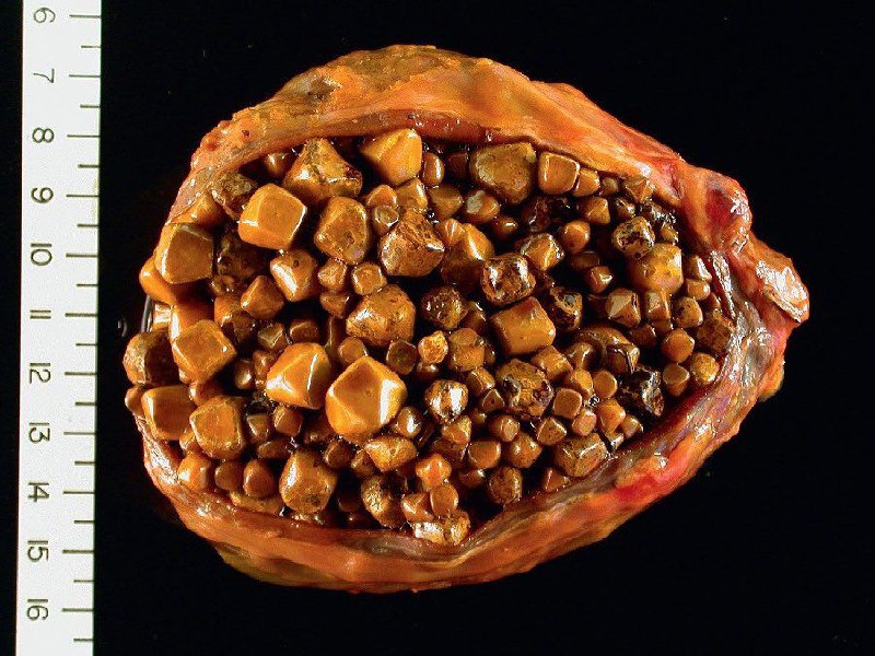
A gallbladder with marked cholelithiasis

A gallstone is a stone formed within the gallbladder out of precipitated bile components. Gallstone disease refers to the condition where gallstones are either in the gallbladder or common bile duct. The presence of stones in the gallbladder is referred to as cholelithiasis.

https://t.me/joinchat/AAAAAEIMp_P_-52SlidzHg
Post #1742 4.01K
Aplasia cutis congenita is a condition in which there is congenital (present from birth) absence of skin, with or without the absence of underlying structures such as bone. It most commonly affects the scalp, but any location of the body can be affected. While most people with aplasia cutis congenita have no other abnormalities, some people have congenital malformations involving the cardiovascular (heart), gastrointestinal, genitourinary, and central nervous systems. The cause of this condition is unclear and appears to be multifactorial; contributing factors may include teratogens, genes, trauma, and compromised blood flow to the skin.

https://t.me/joinchat/AAAAAEIMp_P_-52SlidzHg
Post #1741 3.53K
This is the gross appearance for multicystic renal dysplasia. Note the large but irregular cysts and the asymmetry of the kidneys. The small ureters lead to a hypoplastic bladder.

Multicystic renal dysplasia is a condition that results from the malformation of the kidney during fetal development. The kidney consists of irregular cysts of varying sizes. Multicystic dysplastic kidney is a common type of renal cystic disease, and it is a cause of an abdominal mass in infants.
When a diagnosis of multicystic kidney is made in utero by ultrasound, the disease is found to be bilateral in many cases. Those with bilateral disease often have other severe deformities or polysystemic malformation syndromes.

https://t.me/joinchat/AAAAAEIMp_P_-52SlidzHg
Post #1737 3.86K
A patient was admitted to hospital with green color urine

He explained that he had accidentally ingested the methylene blue dye, a day before was admitted to hospital. The patient was observed over the next 2 days, and the greenish color of urine had disappeared.

https://t.me/joinchat/AAAAAEIMp_P_-52SlidzHg
Older posts →
Threads Profile ViewerView any public Threads profile without an account.Open ThreadLook →Writing with AI? Make it sound human.Metric37 rewrites AI drafts so they read naturally. Free AI detector, 1,500 words free.Try Metric37 →