The ECG demonstrates grouped beats. P-waves precede the QRS complexes with constant but prolonged PR intervals. This indicates a first-degree AV block. Please note that P-wave is absent during the pause between the grouped beats. This pattern suggests an SA node block, rather than a second-degree AV block, in which a P-wave is present during the pause but is not followed by a QRS complex.
For more advanced users: The P-P interval progressively shortens prior to the dropped P wave. The duration of the PP interval during the pause is less than twice the length of the shortest PP interval which is observed prior to the pause. This suggests a second-degree, Wenckebach sinoatrial block.
Special thanks to Dr. Salvatore for his expert ECG interpretation and the informative diagram he provided.
The ECG reveals an irregular bradycardic arrhythmia, characterized by conduction abnormalities at the level of both SA node, as evidenced by the absence of some P waves, and the AV node, where the P waves fail to conduct to the ventricles to produce QRS complexes.
The coexistence of mixed conduction abnormalities in the context of end-stage renal disease (ESRD) raises suspicion for electrolyte imbalances as the primary etiology. This hypothesis was substantiated by laboratory findings indicating elevated levels of potassium (6.7 mg/dL) and decreased levels of calcium (7.1 mg/dL).
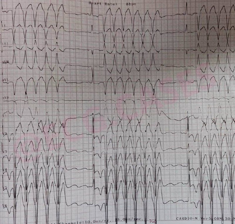
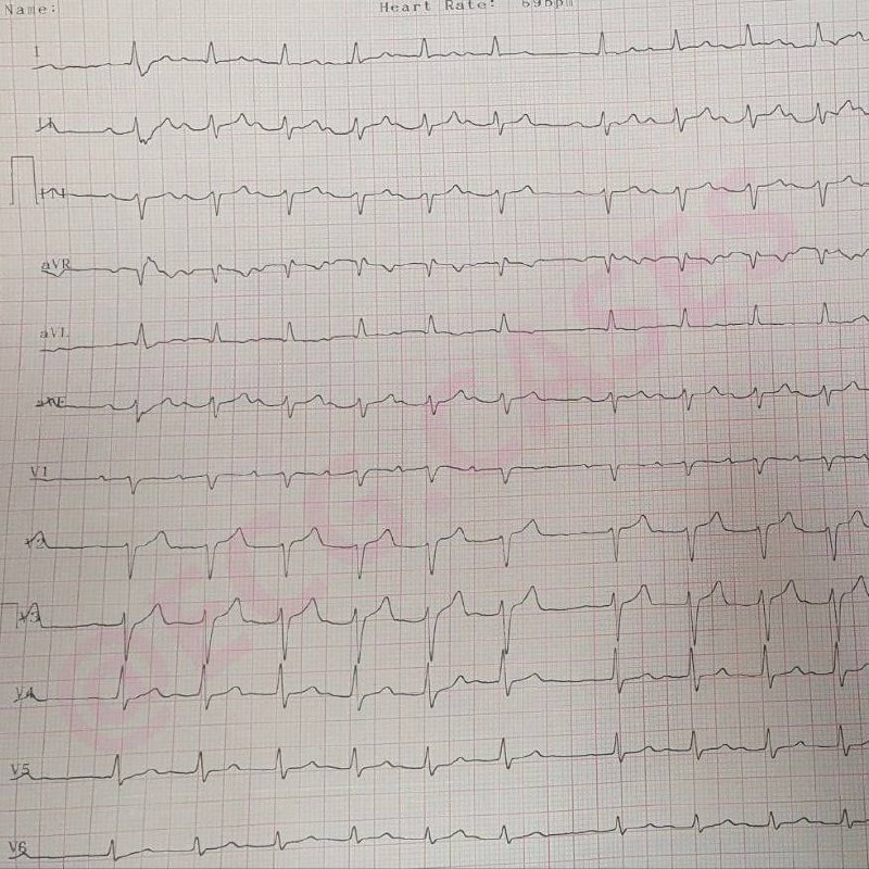
Regular wide complex tachycardias can be either ventricular (VT) or supraventricular (SVT with aberrancy) in origin, and differentiation between the two is usually challenging. Since QRS morphologies in the sinus rhythm are the same as in the broad complex tachycardia, an SVT with aberrancy is more likely than VT.
43-year-old female with history of unknown heart surgery in the childhood presented with sudden onset palpitation and confusion. Due to hemodynamic instability, cardioversion shock was performed and successfully restored its sinus rhythm, which is shown in the next slide.
 EC
EC