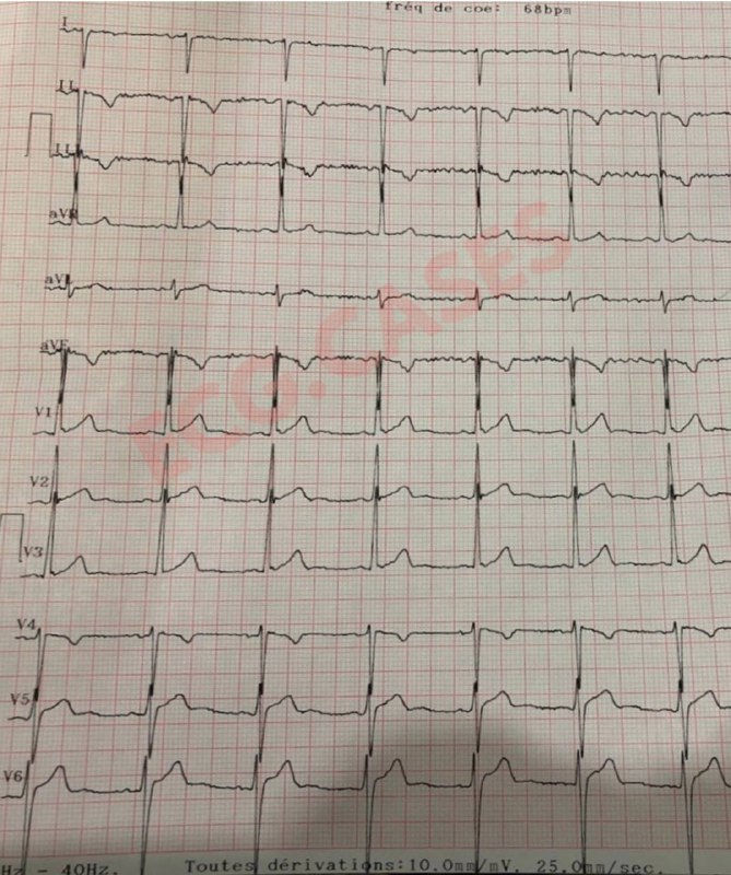
The ECG shows non-conducted PACs, which can sometimes mimic AV block. After each QRS complex, there are premature P waves that appear earlier than expected. These P waves have a different morphology compared to sinus P waves. This difference is most noticeable in V2-V4. Because they occur when the AV node is still in refractory period, they fail to conduct, leading to a pause that can be mistaken for an AV Block.
The ECG shows a regular wide complex tachycardia with a rate of 115 bpm.
Key findings: ✔️ Extreme axis deviation (northwest axis): QRS is positive in aVR and negative in leads I and aVF, which suggests ventricular tachycardia (VT).
In this ECG, the P waves are not clearly visible in most leads because they merged with the T waves. This can sometimes make it tricky to differentiate sinus tachycardia from other arrhythmias. However, if you look closely at lead aVL, you can see a distinct P wave, confirming that the rhythm is sinus. When P waves aren’t obvious, always check multiple leads before ruling out sinus rhythm. Looking at different angles can make all the difference!
 EC
EC